
+917838994961

Currently it only shows your basic business info. Start adding relevant business details such as description, images and products or services to gain your customers attention by using Boost 360 android app / iOS App / web portal.

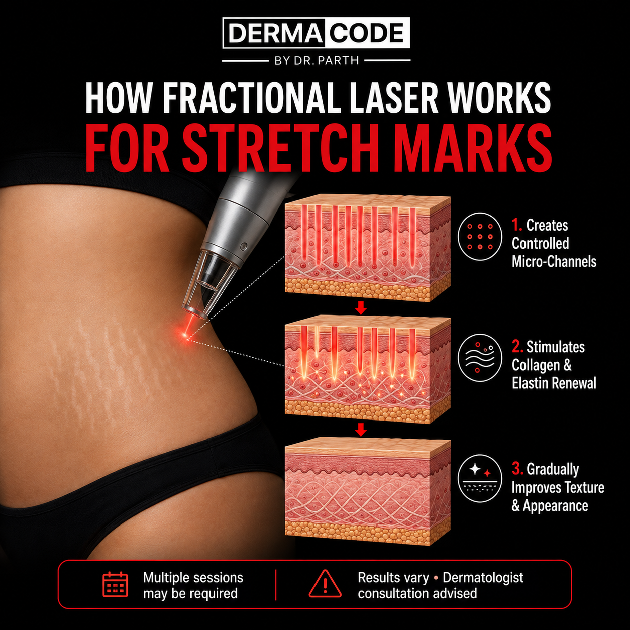
Dermal Remodeling and Tissue Regeneration: How Fractional Laser Works on Stretch Marks Stretch marks, clinically known as striae distensae, are deep dermal scars that develop when the skin undergoes rapid mechanical stretching beyond its biological elasticity—most frequently during pregnancy, adolescent growth spurts, rapid weight fluctuations, or intensive weight training. Under this extreme tension, the structural extracellular matrix located in the reticular dermis ruptures; specifically, bundled Type I and Type III collagen fibers fracture, and the surrounding elastic fiber network fragments, resulting in linear depressions where the overlying epidermis becomes atrophic, thin, and crinkled. In their early vascular stage, known as striae rubrae, these bands appear pink, red, or violaceous due to local capillary dilation and mild inflammation, whereas mature stretch marks, known as striae albae, progress into pale, white, hypo-pigmented, scar-like streaks where blood supply has diminished and structural collagen is largely absent. Because stretch marks represent a profound structural tear situated deep within the mid-to-deep dermal layers rather than a superficial epidermal flaw, standard cosmetic moisturizers, body oils, and topical retinoids struggle to induce noticeable remodeling on their own. Fractional laser therapy—utilizing both ablative Fractional Carbon Dioxide (CO 2 , 10, 600 nm) and non-ablative Erbium-based (such as 1550 nm or 2940 nm) systems—functions on the advanced scientific principle of fractional photothermolysis. Rather than stripping or vaporizing the entire skin surface in a uniform sheet, the fractional scanner breaks the laser beam into an array of microscopic, high-energy laser columns that penetrate through the thinned epidermis down into the damaged reticular dermis. Each micro-beam creates a controlled thermal injury column known as a Microscopic Treatment Zone (MTZ). In ablative fractional lasers, the laser energy vaporizes microscopic plugs of scarred, fractured collagen tissue, creating an open micro-conduit, whereas in non-ablative lasers, intense thermal coagulative zones are generated without outer epidermal vaporization. The defining clinical breakthrough of the fractional approach is that healthy, intact tissue reservoirs are intentionally left untouched between each laser column; these unaffected dermal cells act as biological healing reservoirs that rapidly migrate into the micro-injured zones, dramatically accelerating cellular re-epithelialization while minimizing downtime and procedural risk. The physiological healing cascade triggered within these Microscopic Treatment Zones is what drives true structural repair of stretch marks from the inside out. The controlled micro-thermal damage immediately causes the denatured, loose collagen remnants within the stretch mark base to contract, tightening the lax tissue. Within hours, the body initiates a robust, three-phase wound-healing cascade: inflammatory signaling molecules, growth factors (such as TGF-beta and PDGF), and fibroblasts are recruited to the treated micro-channels. Activated dermal fibroblasts initiate neo-collagenesis (the synthesis of healthy, organized Type I and Type III collagen) and elastogenesis (the regeneration of functional elastin fibers) over a period of several months. As newly synthesized collagen fills the micro-columns, the sunken base of the stretch mark is steadily lifted and reinforced from beneath, closing the textural gap between the scar tissue and the surrounding healthy skin. Simultaneously, fractional laser resurfacing addresses the color discrepancies, lack of microcirculation, and thin texture that make stretch marks so visible to the eye. The laser-induced thermal micro-channels stimulate angiogenesis—the development of new micro-capillaries within previously pale, avascular striae albae—bringing vital nutrients and oxygen back to the scar bed and helping restore natural skin tones. Furthermore, the rapid epidermal turnover induced by the thermal columns sheds dull, atrophic keratinocytes, replacing the thin, papery surface of the stretch mark with fresh, plump epidermal tissue that matches the texture, smoothness, and light-reflective qualities of adjacent skin. When administered through tailored clinical protocols and calibrated stack settings, fractional laser resurfacing systematically softens rigid scar borders, improves skin elasticity, tightens tissue laxity, and blends long-standing stretch marks back into harmonious, healthy skin.


